You can be assured you’re receiving superior care when your healthcare provider orders Imaging Services from Virginia Gay Hospital. Offering MRI, CT, Ultrasound, Bone Densitometry, 3-D Mammography & X-rays.
Confidence and satisfaction comes from understanding that VGH Imaging Services possesses the same advanced, state-of-the art imaging equipment, and industry standard facilities as other hospitals in eastern Iowa, such as those in Cedar Rapids and Iowa City.
Expect prompt appointments, friendly and knowledgeable technologists, and the exact same quality of expertly trained radiologists used by the area’s largest hospitals.
Magnetic Resonance Imaging (MRI) MRI uses a large magnet and radio frequency to align cells in your body to be detected by a scanner. An MRI provides contrast between soft tissue, tendons and ligaments. They can be used for a variety of reasons to detect what is happening within a person’s body. Virginia Gay Hospitaloffers brain and spine MRI’s, sports injury MRI’s and more. Or if you need a specific area such as your shoulders, knees, feet, ankles, neck, cervical and thoracic area checked Viginia Gay Hospital can help. Our hospital has the equipment and specialists needed to diagnose and treat a wide variety of medical conditions.
Our staff understands that MRI scans can be stressful for patients—especially those who experience claustrophobia or discomfort in tight spaces. VGH is proud to offer MRI technology that is designed with comfort in mind. Our MRI machine features a larger, more open design that provides:
More space to reduce feelings of confinement
Greater comfort for patients of all sizes
Less noise and a more relaxed experience
Faster imaging with the same high-quality results your doctor needs
Whether it’s your first scan or a follow-up, our goal is to make the process as easy and stress-free as possible.
Comfort matters. Peace of mind matters. Let VGH help you feel more at ease during your MRI.
To learn more about MRI scan scheduling near me visit our hospital or one of our four conveniently located clinics. The next time your physician orders an MRI, remember VGH for your Vinton MRI Department.
Computed Tomography (CT) is a medical imaging procedure that uses computer-processed X-rays to produce tomographic images or ‘slices’ of specific areas of the body. The cross-sectional images are used for diagnostic and therapeutic purposes. Digital geometry processing is used to generate a three-dimensional image of the inside of an object from a large series of two-dimensional X-ray images taken around a single axis of rotation.
CT scans can be used for a variety of purposes. Whether you need a chest CT for lung health, a CT for cancer screening, a lung cancer screening, a CT for abdominal and pelvic conditions or many other reasons, VGH can help.
CT results are sent to the provider who ordered the test for review. The provider will review the scan and discuss what they did or did not find when they talk to the patient about the next steps.
Next time you need fast diagnostic imaging done, think Virginia Gay Hospital. VGH offers a variety of non-invasive imaging services close to home. To learn more about a CT scan near me, talk to your provider about going to Virginia Gay Hospital for your next scan.
Mammography is the process of using low-energy X-rays to examine the human breast and is used as a diagnostic and a screening tool.
You will now have the ability to schedule your own mammogram at Virginia Gay Hospital. Once an order is placed for that service, you will receive a notice on My Chart and it will ask if you would like to schedule this exam. Choose Virginia Gay Hospital and just follow the prompts, quick and easy instructions, and you’ll be on our schedule. Click on link for more information: Mammogram Self Schedule October 2024
During a mammogram, breasts are compressed to reduce the amount of radiation required to penetrate the tissue. The goal is early detection of breast cancer typically found through characteristic masses and/or microcalcifications. A 3D mammogram screening is available at Virginia Gay Hospital. Research has shown that 3D mammograms in conjunction with standard 2D mammograms detect more cases of invasive breast cancer, while reducing the need for additional testing. The improved results from a 3D mammogram don’t result in radiation exposure above FDA limits. 3D mammograms require only a few seconds more than a standard mammogram. The 3D procedure is identical to a 2D exam, except for the slight arc the imaging unit makes to gather the image. The powerful software creates images of the breast tissue in millimeter-thick increments. The ability to view small segments of tissue and to rotate 3D images allow radiologists to take a more accurate look inside the breast. The 3D technology is especially useful for women with dense breast tissue because 2D mammograms of dense breasts are more difficult to interpret. Breast tissue density is determined after examining a mammogram. Approximately 40% of women have dense breast tissue. Two organizations with information about breast tissue density can be found on the web at http://www.densebreast-info.org/ and http://www.iowabreastdensity.com/.
Talk with your healthcare provider about your risk for breast cancer and their recommendations for a mammogram based on your age and health history. Your provider can provide you with an order for the test. Once you have an order, call the imaging department at 319-472-6288 or scheduling at 319-472-6270 for an appointment. Screening mammograms are scheduled Monday’s from 8:30am-3pm and Tuesday through Friday 8am-3pm. Walk-ins are welcome every day! If you need any financial assistance, the Gifts of Hope fund was established to provide free mammograms and diagnostic services for those in need. Assistance is available to pay for clinic visits, testing, co-pays and diagnostic analysis to determine the next steps, as well as pap tests and/or pelvic exams. Funds are meant for people who have no insurance, have a health insurance policy that does not pay for these services and those who cannot pay for the deductible or co-insurance. Talk to your VGH provider if you want to use the Gifts of Hope fund and arrangements will be made. No application is needed. For more information, visit www.myvghfoundation.org/gifts-of-hope. To learn more about mammogram appointments near me reach out to your healthcare provider or visit one of our four convenient clinics or our hospital.
October is Breast Cancer Awareness month. In October we frequently offer after-hour walk- in mammograms to help patients in our community get the care they need. Our goal is to ease access to mammograms and breast care by offering October Breast Cancer awareness screenings. Take control of your health with Virginia Gay Hospital.”
Accreditation Frequently Asked Questions
What should I know about radiation safety?
Before your imaging procedure be sure to ask your physician the following questions:
Why is the test needed?
How will having the test improve my care?
Are there alternatives that do not use radiation and deliver similar results?
Is the facility accredited by the American College of Radiology (ACR)?
Are pediatric, and adult tests delivered using the appropriate radiation doses?
Why should I have my imaging exam done at an accredited facility?
When you see the gold seals of accreditation prominently displayed in our imaging facility, you can be sure that you are in a facility that meets standards for imaging quality and safety. Look for the ACR Gold Seals of Accreditation.
To achieve the ACR Gold Standard of Accreditation, our facility’s personnel qualifications, equipment requirements, quality assurance, and quality control procedures have gone through a rigorous review process and have met specific qualifications. It’s important for patients to know that every aspect of the ACR accreditation process is overseen by board-certified, expert radiologists, and medical physicists in advanced diagnostic imaging.
What does ACR accreditation mean?
Our facility has voluntarily gone through a vigorous review process to ensure that we meet nationally-accepted standards of care.
Our personnel is well qualified, through education and certification, to perform medical imaging, interpret your images, and administer your radiation therapy treatments.
Our equipment is appropriate for the test or treatment you will receive, and our facility meets or exceeds quality assurance and safety guidelines.
What does the gold seal mean?
When you see the ACR gold seal, you can rest assured that your prescribed imaging test will be done at a facility that has met the highest level of imaging quality and radiation safety. The facility and its personnel have gone through a comprehensive review to earn accreditation status by the American College of Radiology (ACR), the largest and oldest imaging accrediting body in the U.S. and a professional organization of 34,000 physicians.
Nuclear Medicine specialists use safe, painless, and cost-effective techniques to image the body and treat disease. Nuclear medicine imaging is unique, because it provides doctors with information about both structure and function. It is a way to gather medical information that would otherwise be unavailable, require surgery, or necessitate more expensive diagnostic tests. Nuclear medicine imaging procedures often identify abnormalities very early in the progress of a disease—long before many medical problems are apparent with other diagnostic tests.
An Ultrasound is a medical imaging technique used to visualize muscles, tendons and many internal organs. An ultrasound can capture size, structure and pathological lesions with real time images. Ultrasounds can be used to visualize fetuses during routine and emergency prenatal exams. Such diagnostic applications are referred to as obstetric sonography.
Ultrasounds can be used for a variety of reasons including ultrasound-guided procedures. These procedures help a physician get a real time view of what is happening. They can help with accuracy and precision. Many physicians will order an ultrasound for vascular health and an abdominal ultrasound for pain to get a better view and understanding of what is happening with a patient. Ultrasounds can be a great tool to help providers with diagnostics and testing.
If you are interested in learning more about ultrasound imaging at Virginia Gay Hospital talk to your physician or call 319-472-6200.
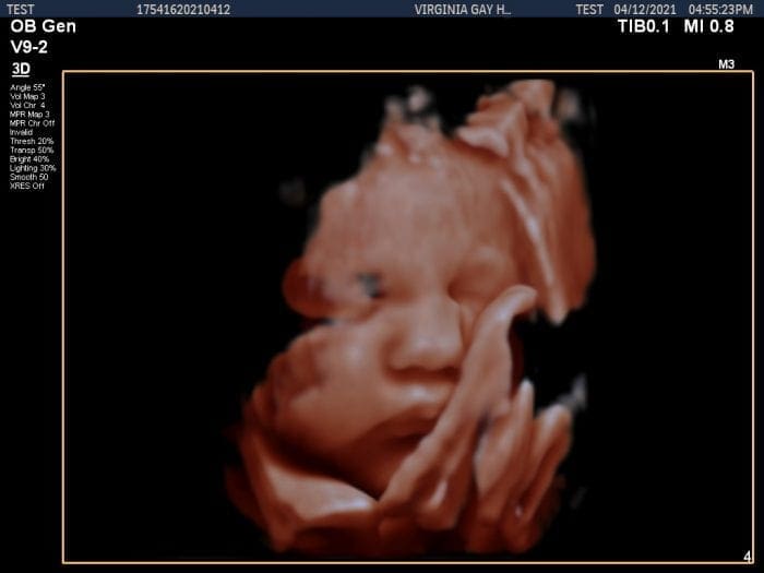
Pregnancy is a monumental time for the child’s parents. Virginia Gay Hospital is excited to offer 3D/4D OB Ultrasounds to commemorate this important event. These 30-minute sessions, located at the main hospital in Vinton, provide high-quality images of your child between 24-36 weeks. Up to three people are allowed in the room during the session. The session costs $125, which must be paid prior to scheduling. After the session, the mother will receive several prints and a thumb drive of all the photos.
To schedule, call 319-472-6288. Please note that these are non-medical/non-diagnostic exams that are not covered by insurance. A radiologist will not read the ultrasounds. Mothers are required to have a normal fetal survey ultrasound at 20 weeks prior to the 3D/4D OB Ultrasound”
Pregnancy is a monumental time for parents. That is why Virginia Gay Hospital offers 3D/4D OB Ultrasounds to commemorate this important time. Our 3D/4D OB Ultrasound sessions are 30-minutes long in the main hospital in Vinton. The Ultrasounds provide high-quality images of your child between 24-36 weeks. Up to three people are allowed in the room during the session and each session costs $125, which must be paid prior to scheduling. After the session, the mother will receive several prints and a thumb drive of all the photos.
If you are interested in scheduling a 3D/4D OB Ultrasound, call 319-472-6288. Please note that these are non-medical/non-diagnostic exams that are not covered by insurance. A radiologist will not read the ultrasounds. Mothers are required to have a normal fetal survey ultrasound at 20 weeks prior to a 3D/4D OB Ultrasound.
If you are looking for a prenatal ultrasound near me or other affordable ultrasound imaging, reach out to your Obstetrician to schedule an appointment close to home at at Virginia Gay Hospital.
Providing high-quality services to commemorate your pregnancy.
Pregnancy is a monumental time for the child’s parents. Virginia Gay Hospital is excited to offer 3D/4D OB Ultrasounds to commemorate this important event. These 30-minute sessions, located at the main hospital in Vinton, provide high-quality images of your child between 24-36 weeks. Up to three people are allowed in the room during the session. The session costs $125, which must be paid prior to scheduling. After the session, the mother will receive several prints and a thumb drive of all the photos.
To schedule, call 319-472-6288. Please note that these are non-medical/non-diagnostic exams that are not covered by insurance. A radiologist will not read the ultrasounds. Mothers are required to have a normal fetal survey ultrasound at 20 weeks prior to the 3D/4D OB Ultrasound
An X-ray is an imaging test that uses small doses of radiation to show internal organs and bones. X-rays are used to diagnose fractures, fluid buildup, infections and much more. They are a form of electromagnetic radiation that can penetrate clothing, body tissue and internal organs. An X-ray machine sends radiation through a patient’s body to create an x-ray image. Radiation is absorbed differently throughout the body, so the machine will create a black and white image of the patients’ internal structures based on how much radiation is absorbed and where. The patient’s provider is sent a copy of the images to review, diagnose and treat as needed.
If you are looking for an affordable x-ray or an x-ray walk-in clinic close to home, choose Virginia Gay Hospital. Our imaging department makes sure that patients feel comfortable and relaxed. We take the worry out of healthcare. Whether you need a digital x-ray service, a broken bone x-ray or sports injury imaging, Virginia Gay Hospital is here for you
Bone Densitometry A bone density test is a non-surgical, painless examination that uses low dose x-rays to measure the density of bones. A bone density test tells you if you have normal bone density, low bone density (osteopenia) or osteoporosis. It is the only test that can diagnose osteoporosis. The lower your bone density, the greater your risk of breaking a bone.
Virginia Gay Hospital and Clinics created this web page for information about the COVID-19 virus and responses by VGH to provide care to patients and the communities we serve.
Please call ahead if you are experiencing symptoms of the flu or COVID-19.


 Providing high-quality services to commemorate your pregnancy.